Скачать с ютуб Testicular Hydrocele, pyocele, varicocele, and Inguinal Hernia. в хорошем качестве
Testicular Hydrocele, pyocele, varicocele, and Inguinal Hernia.
3 года назад
Скачать бесплатно и смотреть ютуб-видео без блокировок Testicular Hydrocele, pyocele, varicocele, and Inguinal Hernia. в качестве 4к (2к / 1080p)
У нас вы можете посмотреть бесплатно Testicular Hydrocele, pyocele, varicocele, and Inguinal Hernia. или скачать в максимальном доступном качестве, которое было загружено на ютуб. Для скачивания выберите вариант из формы ниже:
Загрузить музыку / рингтон Testicular Hydrocele, pyocele, varicocele, and Inguinal Hernia. в формате MP3:
Если кнопки скачивания не
загрузились
НАЖМИТЕ ЗДЕСЬ или обновите страницу
Если возникают проблемы со скачиванием, пожалуйста напишите в поддержку по адресу внизу
страницы.
Спасибо за использование сервиса savevideohd.ru
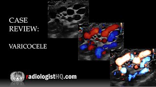
Testicular Hydrocele, pyocele, varicocele, and Inguinal Hernia.
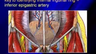
This video shows Testicular Hydrocele, pyocele, varicocele, and Inguinal Hernia. Ultrasound is the imaging modality of choice for the investigation of scrotal pain and swelling or follow-up of a known epididymo-orchitis. Features of pyocele on ultrasound are complex, heterogeneous fluid collection in the scrotal sac with septa. Gas may be present causing hyperechoic foci and shadowing. At sonography, congenital hydrocele appears as an anechoic fluid collection surrounding the anterolateral aspects of the testis and sometimes extending to the inguinal canal or as a fluid collection with low-level swirling echoes, which are related to protein aggregation or deposition of cholesterol crystals. A hydrocele can develop as a result of injury or inflammation within the scrotum. Inflammation might be caused by an infection in the testicle or in the small, coiled tube at the back of each testicle (epididymitis). A hydrocele is defined as fluid surrounding the testicle, which is usually simple, anechoic, and without layering debris. If septations, significant layering, or debris are visualized during scanning, then the diagnosis of a complex hydrocele is made and either pyocele or hematocele is considered. Varicoceles are abnormal dilatations of the pampiniform venous plexus. They are classified as primary or secondary, depending on their cause, and staged clinically on the basis of their extension and on the presence or the absence of spontaneous or induced reversal of blood flow. Signs of varicoceles on ultrasound are veins that are wider than 3 millimeters with blood flowing the wrong way during the Valsalva maneuver. The ultrasound can also show the size of the testicles. Two types can be distinguished on the basis of etiopathogenesis: primary and secondary. Primary varicoceles are due to venous reflux into the pampiniform plexus from the internal spermatic vein as a result of incontinent venous valves, and they usually occur on the left side (85% of cases). In fact, the left internal spermatic vein drains into the homolateral renal vein at a 90-degree angle, and as a result of this particular anatomical feature, endoluminal pressure in the renal vein is transmitted backward, opposing flow from the internal spermatic vein. On the right side, the internal spermatic vein merges with the inferior cava vein at an acute angle, and its drainage is promoted by the negative pressure created by caval blood flow. Secondary varicoceles are the result of increased pressure in the testicular vein, which can be caused by several conditions, such as hydronephrosis, hepatic cirrhosis associated with portal hypertension with splenorenal shunts, abdominal and retroperitoneal neoplasms, and the so-called nutcracker phenomenon, which involves compression of the left renal vein between the superior mesenteric artery and aorta. A hernia occurs when an organ or fatty tissue squeezes through a weak spot in a surrounding muscle or connective tissue called fascia. The most common types of hernia are inguinal (inner groin), incisional (resulting from an incision), femoral (outer groin), umbilical (belly button), and hiatal (upper stomach). An inguinal hernia occurs when tissue, such as part of the intestine, protrudes through a weak spot in the abdominal muscles. The resulting bulge can be painful, especially when you cough, bend over or lift a heavy object. Ultrasound is a non-invasive, non-ionizing radiation modality that is highly successful at soft tissue imaging. Groin pain from an occult hernia can be a difficult clinical diagnosis made easier by good imaging by optimizing the image using depth, focus, and gain. Ultrasound findings in Inguinal Hernia 1). Herniated gut loops along with some fluid collection at the Inguinal region. 2). Testis and epididymis pulled up with herniated contents. 3). Hydrocele on the respective involved side. 4). Normal testis on the unaffected side. A bulge in the groin area is visible. Because standing and coughing can make a hernia more prominent, you'll likely be asked to stand and cough or strain. If the diagnosis isn't readily apparent, your doctor might order an imaging test, such as an abdominal ultrasound, CT scan, or MRI. Examination of an adult for an inguinal hernia is best performed from the seated position, with the patient standing. The inguinal canal areas for the bulge are visualized. A provocative cough may be necessary to expose the hernia; the cough is repeated as the examiner invaginates the scrotum and feels for an impulse. Not all inguinal hernias need to be repaired, but all hernia repairs require surgery. Small hernias that are not strangulated, not blocking the blood supply to the intestine, and are not causing bowel obstruction or significant pain do not necessarily require surgery or emergency surgical repair. An inguinal hernia isn't necessarily dangerous. But it doesn't improve on its own and can lead to life-threatening complications.
Comments