Скачать с ютуб Histology of the urinary system в хорошем качестве
Histology of the urinary system
1 год назад
Скачать бесплатно и смотреть ютуб-видео без блокировок Histology of the urinary system в качестве 4к (2к / 1080p)
У нас вы можете посмотреть бесплатно Histology of the urinary system или скачать в максимальном доступном качестве, которое было загружено на ютуб. Для скачивания выберите вариант из формы ниже:
Загрузить музыку / рингтон Histology of the urinary system в формате MP3:
Если кнопки скачивания не
загрузились
НАЖМИТЕ ЗДЕСЬ или обновите страницу
Если возникают проблемы со скачиванием, пожалуйста напишите в поддержку по адресу внизу
страницы.
Спасибо за использование сервиса savevideohd.ru
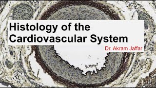
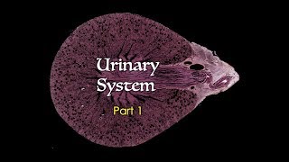
Histology of the urinary system
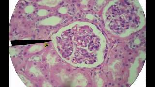
By the end of this video, you should be able to Differentiate between Renal cortex and medulla. Proximal convoluted tubule, distal convoluted tubule, collecting duct, thick limb of the loop of Henle, thin limb of the loop of Henle, and capillary lumen. The glomerulus, Bowman’s capsule, Bowman’s space, and renal corpuscle. Renal pole and vascular pole of the Bowman’s capsule. Identify: The layers of the wall of the urinary bladder The layers of the wall of the ureter Transitional epithelium Juxtaglomerular apparatus, basement membrane, medullary ray, mesothelium, minor calyx, renal papilla. Describe The function of the transitional epithelium. The functional unit of the kidney and the distribution of its components. Explain The presence of a brush border and dark cytoplasm in proximal convoluted tubules. Explain the abundance of profiles of proximal convoluted tubules in comparison to the distal. Presented and edited by Dr. Akram Jaffar, Ph.D. This video and its channel are supported by the "Human Anatomy Education" Page on Facebook / anatomyeducation Related accounts Twitter / akramjaffar Facebook / anatomyeducation SlideShare http://www.slideshare.net/AkramJaffar LinkedIn / akram-abo. . Research gate https://www.researchgate.net/profile/... Medtube https://medtube.net/users/akram-jaffar Instagram / akramjaffar Academia https://dal.academia.edu/AkramJaffar
Comments