РЎРәР°СҮР°СӮСҢ СҒ СҺСӮСғРұ Knee Joint Anatomy Animation : Bones, Ligaments, Menisciп»ҝ, Innervation, Blood supply and Movements РІ С…РҫСҖРҫСҲРөРј РәР°СҮРөСҒСӮРІРө
Knee Joint Anatomy Animation : Bones, Ligaments, Menisciп»ҝ, Innervation, Blood supply and Movements
11 РјРөСҒСҸСҶРөРІ РҪазаРҙ
РЎРәР°СҮР°СӮСҢ РұРөСҒРҝлаСӮРҪРҫ Рё СҒРјРҫСӮСҖРөСӮСҢ СҺСӮСғРұ-РІРёРҙРөРҫ РұРөР· РұР»РҫРәРёСҖРҫРІРҫРә Knee Joint Anatomy Animation : Bones, Ligaments, Menisciп»ҝ, Innervation, Blood supply and Movements РІ РәР°СҮРөСҒСӮРІРө 4Рә (2Рә / 1080p)
РЈ РҪР°СҒ РІСӢ РјРҫР¶РөСӮРө РҝРҫСҒРјРҫСӮСҖРөСӮСҢ РұРөСҒРҝлаСӮРҪРҫ Knee Joint Anatomy Animation : Bones, Ligaments, Menisciп»ҝ, Innervation, Blood supply and Movements или СҒРәР°СҮР°СӮСҢ РІ РјР°РәСҒималСҢРҪРҫРј РҙРҫСҒСӮСғРҝРҪРҫРј РәР°СҮРөСҒСӮРІРө, РәРҫСӮРҫСҖРҫРө РұСӢР»Рҫ загСҖСғР¶РөРҪРҫ РҪР° СҺСӮСғРұ. ДлСҸ СҒРәР°СҮРёРІР°РҪРёСҸ РІСӢРұРөСҖРёСӮРө РІР°СҖРёР°РҪСӮ РёР· С„РҫСҖРјСӢ РҪРёР¶Рө:
ЗагСҖСғР·РёСӮСҢ РјСғР·СӢРәСғ / СҖРёРҪРіСӮРҫРҪ Knee Joint Anatomy Animation : Bones, Ligaments, Menisciп»ҝ, Innervation, Blood supply and Movements РІ С„РҫСҖРјР°СӮРө MP3:
Р•СҒли РәРҪРҫРҝРәРё СҒРәР°СҮРёРІР°РҪРёСҸ РҪРө
загСҖСғзилиСҒСҢ
РқРҗР–РңРҳРўР• ЗДЕСЬ или РҫРұРҪРҫРІРёСӮРө СҒСӮСҖР°РҪРёСҶСғ
Р•СҒли РІРҫР·РҪРёРәР°СҺСӮ РҝСҖРҫРұР»РөРјСӢ СҒРҫ СҒРәР°СҮРёРІР°РҪРёРөРј, РҝРҫжалСғР№СҒСӮР° РҪР°РҝРёСҲРёСӮРө РІ РҝРҫРҙРҙРөСҖР¶РәСғ РҝРҫ Р°РҙСҖРөСҒСғ РІРҪРёР·Сғ
СҒСӮСҖР°РҪРёСҶСӢ.
РЎРҝР°СҒРёРұРҫ Р·Р° РёСҒРҝРҫР»СҢР·РҫРІР°РҪРёРө СҒРөСҖРІРёСҒР° savevideohd.ru
Knee Joint Anatomy Animation : Bones, Ligaments, Menisciп»ҝ, Innervation, Blood supply and Movements
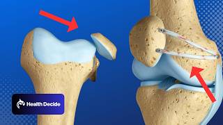
рҹ“Ң рқҗ…рқҗЁрқҗҘрқҗҘрқҗЁрқҗ° рқҗЁрқҗ§ рқҗҲрқҗ§рқҗ¬рқҗӯрқҗҡрқҗ рқҗ«рқҗҡрқҗҰ:- В В /В drgbhanuprakashВ В рҹ“Ңрқ—қрқ—јрқ—¶рқ—» рқ—ўрқҳӮрқ—ҝ рқ—§рқ—Ірқ—№рқ—Ірқ—ҙрқ—ҝрқ—®рқ—ә рқ—–рқ—өрқ—®рқ—»рқ—»рқ—Ірқ—№ рқ—ӣрқ—Ірқ—ҝрқ—І:- https://t.me/bhanuprakashdr рҹ“Ңрқ—ҰрқҳӮрқ—ҜрқҳҖрқ—°рқ—ҝрқ—¶рқ—Ҝрқ—І рқ—§рқ—ј рқ— рқҳҶ рқ— рқ—®рқ—¶рқ—№рқ—¶рқ—»рқ—ҙ рқ—ҹрқ—¶рқҳҖрқҳҒ:- https://linktr.ee/DrGBhanuprakash Knee Joint Anatomy Animation : Ligaments, Menisciп»ҝ, Innervation, Blood supply and Movements The knee joint is one of the most complex and largest joints in the human body, crucial for performing everyday activities like walking, running, and jumping. Comprising various structures including bones, ligaments, tendons, and cartilage, each component plays a vital role in the function and stability of the knee. Bones Involved in the Knee Joint ________________________________ Femur: The thigh bone, or femur, is the longest and strongest bone in the body. Its lower end forms the upper part of the knee joint. Tibia: The shin bone, or tibia, forms the lower part of the knee joint. Patella: Also known as the kneecap, the patella is a small, triangular bone that protects the knee and provides leverage for leg extension. Ligaments and Their Functions ______________________________ Anterior Cruciate Ligament (ACL): One of the most commonly discussed ligaments, the ACL provides rotational stability and prevents the tibia from sliding out in front of the femur. Posterior Cruciate Ligament (PCL): The PCL works oppositely to the ACL, preventing the tibia from sliding backwards under the femur. Medial Collateral Ligament (MCL): Located on the inner side of the knee, the MCL resists forces acting from the outside of the knee. Lateral Collateral Ligament (LCL): Found on the outer side of the knee, the LCL resists forces coming from the inner side of the knee. Cartilage and Cushioning Meniscus: These are crescent-shaped pads of cartilage situated between the femur and tibia that act as shock absorbers and provide stability. Medial Meniscus: Located on the inner side of the knee. Lateral Meniscus: Located on the outer side of the knee. Articular Cartilage: This is the smooth, white tissue covering the ends of bones where they come into contact with each other. It allows for smooth movement within the joint and acts as a cushion between the bones. Tendons and Muscles ______________________ Quadriceps Tendon: This tendon connects the quadriceps muscle to the patella and is essential for straightening the knee. Patellar Tendon: This connects the patella to the tibia and is part of the mechanism that allows for knee extension and flexion. Hamstrings: These muscles are located at the back of the thigh and are responsible for bending the knee. Additional Structures Bursae: These are small sacs filled with fluid that reduce friction between the moving parts of the knee. Synovial Membrane: This lines the joint and secretes synovial fluid for lubrication. #fmge #fmgevideos #rapidrevisionfmge #fmgejan2023 #mbbslectures #nationalexitexam #nationalexittest #neetpg #usmlepreparation #usmlestep1 #fmge #usmle #drgbhanuprakash #medicalstudents #medicalstudent #medicalcollege #neetpg2023 #usmleprep #usmlevideos #usmlestep1videos #medicalstudents #neetpgvideos #kneejoint #knee #kneepain #lowerlimbanatomy #lowerlimb #kneearthritis #kneejointanatomy #kneeanatomy
Comments