Скачать с ютуб General Embryology - Detailed Animation On Second Week Of Development в хорошем качестве
General Embryology - Detailed Animation On Second Week Of Development
10 лет назад
Скачать бесплатно и смотреть ютуб-видео без блокировок General Embryology - Detailed Animation On Second Week Of Development в качестве 4к (2к / 1080p)
У нас вы можете посмотреть бесплатно General Embryology - Detailed Animation On Second Week Of Development или скачать в максимальном доступном качестве, которое было загружено на ютуб. Для скачивания выберите вариант из формы ниже:
Загрузить музыку / рингтон General Embryology - Detailed Animation On Second Week Of Development в формате MP3:
Если кнопки скачивания не
загрузились
НАЖМИТЕ ЗДЕСЬ или обновите страницу
Если возникают проблемы со скачиванием, пожалуйста напишите в поддержку по адресу внизу
страницы.
Спасибо за использование сервиса savevideohd.ru
General Embryology - Detailed Animation On Second Week Of Development
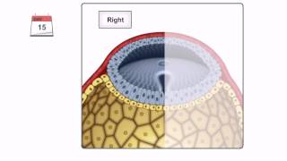
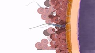
Implantation of the blastocyst usually occurs 6-8 days after fertilization. By day 8, the blastocyst has burrowed into the uterine wall, being completely embedded by day 9. Day 8 The blastocyst is composed of two main components: the outer cell mass, the trophoblast; and the inner cell mass, the embryoblast. As the trophoblast makes contact with the endometrium, it differentiates into two layers: an inner, cytotrophoblast; and an outer, syncytiotrophoblast. The embryoblast differentiates into a bilaminar embryonic disc composed of two cell layers, the hypoblast, and the epiblast. Soon after the embryonic disc has formed, a cavity begins to appear between the epiblast and cytotrophoblast, known as the amniotic cavity. Cells originating from the hypoblast begin to migrate, forming a thin membrane, which lines the inner surface of the cytotrophoblast. This is called the exocoelomic membrane. The exocoelomic membrane and cells of the hypoblast together form the walls of the primitive yolk sac. Day 9 Once the blastocyst is completely embedded in the uterus wall, a plug called a fibrin coagulum forms in the gap created in the epithelium of the uterus by the blastocyst. At this time, the growth of the cytotrophoblast and syncytiotrophoblast is much faster than that of the bilaminar embryonic disc Small holes called lacunae begin to form in the syncytiotrophoblast as it continues to expand. Day 12 Capillaries in the endometrium surrounding the developing embryo dilate, forming maternal sinusoids. As the lacunae continue to expand, enzymes within the syncytiotrophoblast begin to erode the lining of the sinusoids and uterine glands. This allows anastomosis between the maternal sinusoids and lacunar networks to begin establishment of an uteroplacental circulation. By day 12, the lacunae stop growing and fuse to form large interconnecting spaces, called lacunar networks. A new population of cells appears between the inner surface of the cytotrophoblast and the outer surface of the primitive yolk sac, known as the extraembryonic mesoderm. Large cavities begin to appear in the extraembryonic mesoderm. These gradually fuse to form one single cavity, called an extraembryonic coelom. Day 13 Approximately 13 days after fertilization, a large portion of the exocoelomic cavity is pinched off, forming a smaller cavity, called the secondary yolk sac. A remnant also forms within the exocoelomic cavity, known as an exocoelomic cyst, which is eventually eliminated. By the end of the second week of development, the chorionic cavity enlarges, and the bilaminar embryonic disc is joined to the trophoblast by a band of extraembryonic mesoderm called the connecting stalk. __________________________ Join us on this educational journey to expand your knowledge and gain a deeper understanding of the human body, diseases, treatments, and cutting-edge medical advancements. Make sure to "subscribe" to get our new videos. If you enjoy our content and would like to support us, we now accept donations in Bitcoin. Your contributions help us create more awesome videos for you. Consider sending a donation to our Bitcoin address and be a part of the journey! Thank you for your incredible support! Bitcoin: bc1q00nhyd0smnd42jjqjdwsady02x83maatvfla0c ETH: 0xf09e1854063638f4Ed13F42392a84B99b0093AA5
Comments