Скачать с ютуб Detailed anomaly scan в хорошем качестве
Detailed anomaly scan
11 лет назад
Detailed anomaly scan
foetal anatomy scan
fetal anomaly scan
detailed anatomy scan
anatomy scan
anomaly scan
pregnancy
18th week of pregnancy
19th week of pregnancy
20th week of pregnancy
21st week of pregnancy
22nd week of pregnancy
pregnancy screening
pregnancy scan
pregnancy anomaly scan
foetus
foetal anomaly scan
ultrasound scan
ultrasound screening
pregnancy ultrasound
ultrasound
Скачать бесплатно и смотреть ютуб-видео без блокировок Detailed anomaly scan в качестве 4к (2к / 1080p)
У нас вы можете посмотреть бесплатно Detailed anomaly scan или скачать в максимальном доступном качестве, которое было загружено на ютуб. Для скачивания выберите вариант из формы ниже:
Загрузить музыку / рингтон Detailed anomaly scan в формате MP3:
Если кнопки скачивания не
загрузились
НАЖМИТЕ ЗДЕСЬ или обновите страницу
Если возникают проблемы со скачиванием, пожалуйста напишите в поддержку по адресу внизу
страницы.
Спасибо за использование сервиса savevideohd.ru
Detailed anomaly scan
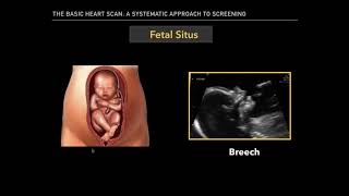
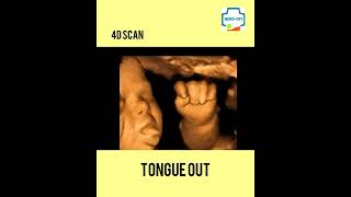
Fetal anatomy scan is one of the most important ultrasound examinations during the pregnancy. This examination is performed with the transabdominal probe during the 18–22 week of gestation. By that time the fetal organs have formed and the period of growth and maturing begins. By the 20th week approximately half of the pregnancy will be over and the other half still ahead. By that time foetus weighs around 300–350 grams and is about 21–22 cm long. The foetus already resembles a human baby; however, it can`t smile as its facial mimic muscles have not yet developed. The foetus is able to open the mouth, it swallows and breaths amniotic liquid and senses its smell and taste. The foetus’s inner ear has developed, so it can hear the sounds coming from the womb: mother’s heartbeat, breathing murmur of lungs, and peristaltic movement of the intestine. However, its not able to distinguish sounds coming from outside the womb, for example father’s speech. The skin is covered with down soft hair and vernix caseosa to protect the foetus from the macerating impact of the amniotic fluid. The foetus has already eyebrows and eyelids have formed, but it can not open the eyes as they are still covered with embryonic membrane. The foetus has no hair or eyelashes yet; however, nails have started to form at the end of the fingers and toes. The foetus’s heart is the size of a 1 euro coin and it beats about 120–160 times a minute or twice as fast as the mother’s heart or the mother’s and father’s heart together. During the ultrasound examination the size and structure of the foetus and the attachment of the umbilical cord in the placenta is examined. It is important to see that the lower edge of the placenta does not cover the neck of the uterus in order not to hinder the labor process. The amount of amniotic water is assessed to make sure there is not to too little or too much of it. The size of the foetal skull, the circumference of the belly and the length of the thighbone are measured to assess the development and the time of labor. It is very important to evaluate the foetal organ structures in detail. Attention should be paid to the skull, which has to be complete, and the seams of the skull and fontanels have to be open so that the growth of the brain would be unhindered. The brain structure and its compliance to the gestation period is evaluated. Attention will be paid to the foetal eyes and the width between the eyes. The upper lip is evaluated so that one born the baby could have a beautiful smile. The lower jaw is assessed so that the baby will be able to latch and suck properly in the future. The typical ultrasound features referring to chromosome diseases are observed. Attention is paid to the completeness of the foetal spine and the front wall of the stomach and the umbilical cord proceeding from the belly. The completeness, position and movement of the baby’s hands and legs are being assessed. The foetus can bend and stretch hands and legs and can open and close its hand. Attention should be paid to the kidneys, their structure and function is assessed, it is observed whether the bladder is filled and whether the arteries of the umbilical cord surround it. You have to observe whether the baby swallows and its stomach is full, and whether it’s properly located on the left side of its body along with the heart. Special attention should be paid to the foetus heart structure, which has to have four chambers. Major blood vessels have to proceed from the right chambers and the heartbeat must be regular. In case of the increased risk of premature labor, it is possible to measure the length of the neck of the uterus by an intra-vaginal probe and estimate the risk of premature birth. During the ultrasound examination the embryo’s gender can also be determined should the parents wish to know it before delivery. After the end of the ultrasound examination the results are explained to the family and the essence of possible deviations, reasons and prognosis are explained in detail. It is important to know that the majority of babies develop well and are born healthy.
Comments