Скачать с ютуб 2-Minute Neuroscience: The Ventricles в хорошем качестве
2-Minute Neuroscience: The Ventricles
9 лет назад
Скачать бесплатно и смотреть ютуб-видео без блокировок 2-Minute Neuroscience: The Ventricles в качестве 4к (2к / 1080p)
У нас вы можете посмотреть бесплатно 2-Minute Neuroscience: The Ventricles или скачать в максимальном доступном качестве, которое было загружено на ютуб. Для скачивания выберите вариант из формы ниже:
Загрузить музыку / рингтон 2-Minute Neuroscience: The Ventricles в формате MP3:
Если кнопки скачивания не
загрузились
НАЖМИТЕ ЗДЕСЬ или обновите страницу
Если возникают проблемы со скачиванием, пожалуйста напишите в поддержку по адресу внизу
страницы.
Спасибо за использование сервиса savevideohd.ru
2-Minute Neuroscience: The Ventricles
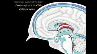
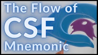
In my 2-Minute Neuroscience videos I explain neuroscience topics in 2 minutes or less. In this video, I cover the ventricles. I discuss the function of the ventricles, which involves production and distribution of cerebrospinal fluid; I also briefly explain the functions of cerebrospinal fluid. I describe the structure of the ventricles, including descriptions of the lateral, third, and fourth ventricles, as well as the means by which the ventricles are connected to one another: the interventricular foramen and cerebral aqueduct. Finally, I mention hydrocephalus, a condition that occurs when cerebrospinal fluid levels in the ventricles get too high. For an article (on my website) that explains the ventricles, click this link: https://neuroscientificallychallenged... TRANSCRIPT: Welcome to 2 minute neuroscience, where I simplistically explain neuroscience topics in 2 minutes or less. In this installment I will discuss the ventricles. The ventricles are a network of cavities that are distributed throughout the brain. They are lined with a specialized membrane called the choroid plexus, which is composed of glial cells called ependymal cells. Ependymal cells are specially designed to secrete cerebrospinal fluid, which then flows through the ventricles and around the brain. Thus the main role of the ventricles is the production and distribution of cerebrospinal fluid. Cerebrospinal fluid, or CSF as it is commonly called, is a clear colorless liquid that performs a number of important functions in the brain. Among other things, csf surrounds the brain, forming a protective layer and suspending the brain in fluid, which reduces the strain forces like gravity would have on the brain. Also, it constantly flows through and around the brain removing toxins and regulating the extracellular environment of neurons. There are 4 ventricles. There are two C-shaped lateral ventricles, one in each of the cerebral hemispheres. The lateral ventricles are connected to a third ventricle by an opening called the interventricular foramen. The third ventricle is a narrow cavity that runs along the midline of the diencephalon. It looks something like a misshapen donut, and the section that would be the hole of the donut is an area known as the interthalamic adhesion. In this section the thalamus makes up the wall of the third ventricle. The third ventricle communicates with the fourth ventricle via the cerebral aqueduct. The fourth ventricle is wedged between the cerebellum on one side and the brainstem on the other. The fourth ventricle is shaped like a tent whose peak protrudes into the cerebellum. There are three openings in the fourth ventricle that allow cerebrospinal fluid to enter the subarachnoid space, a CSF-containing cavity that surrounds the brain. Thus, CSF leaves the ventricular system via the 4th ventricle. The 4th ventricle extends to, and is continuous with, the central canal, a CSF-filled cavity that runs the length of the spinal cord. If CSF circulation is blocked as by a tumor, or is otherwise abnormally excessive, it can result in increased pressure and expansion of the ventricles. This creates a condition known as hydrocephalus, which can cause a variety of complications and be life-threatening. REFERENCE: Nolte J. The Human Brain: An Introduction to its Functional Anatomy. 6th ed. Philadelphia, PA. Elsevier; 2009.
Comments